Examen échographique connaissances scientifiques populaires
——The difference between "B ultrasound" and "color ultrasound"
During daily ultrasound examinations, we found that many patients would ask, "Doctor, are you a color ultrasound? Why is it black and white?". Commonly known as "B-ultrasound", it uses the probe to scan and repeatedly transmit and receive ultrasonic sound beams to form a two-dimensional cross-sectional image displayed on the screen, that is, two-dimensional ultrasound. Gray-scale cross-sectional images of organ structure and morphology.
L'imagerie par ultrasons utilise des changements d'échelle de gris du blanc au gris au noir pour afficher les changements dans les propriétés physiques des organes ou des lésions, tels que les liquides apparaissant en noir, les calculs biliaires, les os ou les gaz apparaissant en blanc brillant ; en raison de la vitesse d'imagerie par ultrasons rapide, il peut afficher la situation-en temps réel de l'activité des organes ; l'échographie bidimensionnelle-a une orientation : les images en niveaux de gris-des tranches de l'axe XY du haut, du bas, de la gauche, de la droite, de l'avant et de l'arrière, et l'axe Z nécessite trois- balayage dimensionnel.
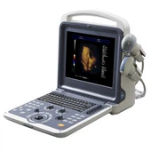
With the gradual improvement of medical conditions in our country, many larger medical institutions have already eliminated this simple two-dimensional ultrasound instrument and replaced it with a color Doppler ultrasound examination system. People call it "color ultrasound". The actual color ultrasound instrument has abdominal Color Doppler actually includes B-ultrasound, color Doppler, M-mode echocardiography and other related inspection and analysis software. The function and scope of application are related to the specific configuration of the instrument. The functions of black and white ultrasound and color ultrasound are distinguished.
Simply put, "color Doppler" is a high-definition black and white B-ultrasound plus color Doppler. Its main advantages are: it can quickly and intuitively display the two-dimensional plane distribution of blood flow; it can display the running direction of blood flow; it is beneficial to distinguish between arteries and veins; it is beneficial to identify vascular lesions and non-vascular lesions; it is beneficial to understand the nature of blood flow It can easily understand the time phase and speed of blood flow; can reliably find shunt and reflux; can quantitatively analyze the origin, width, length and area of blood flow bundles. These ultrasound manifestations and parameters play a very important auxiliary role in the diagnosis of clinical diseases.
"Color Doppler" can display the changes of blood perfusion in organs or lesions, distinguish solid or cystic masses, and prompt according to the changes of the blood flow spectrum of the cyst wall and solid masses, such as blood flow velocity, shape, resistance, and blood perfusion characteristics The reference basis for qualitative diagnosis, such as low-resistance solid masses are more common in malignant tumors.
"Color Doppler" to determine the hemodynamic changes of the cardiovascular system: filling state, blood flow color, direction, path, speed, pressure, phase, time, flow, etc. for disease diagnosis; such as small atrioventricular septal defects, two-dimensional Ultrasound may not show the gap, but once the color Doppler is on, it can clearly show the shunt of the defect site, which plays an important role in auxiliary diagnosis; another example is heart valve stenosis and vascular stenosis, when blood flow passes through the stenosis. , Due to the increase in proximal pressure, multicolored mosaic jets will appear in the stenotic orifice and the distal end. Doctors can quantify the valve area by measuring the half-time of pressure drop by spectral Doppler.
Therefore, the difference between "color ultrasound" and "B ultrasound" cannot be equal to the difference between color TV and black and white TV.